PET/CT – innovative diagnostic advance
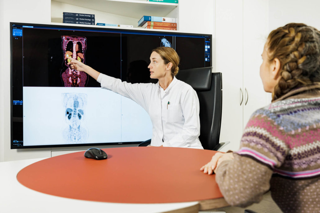
PET/CT plays an important role in the fight against cancer today. The combination of PET and CT brings enormous advantages over positron emission tomography (PET) alone. While PET is used to record metabolic processes, computed tomography makes the function of the examined organism visible.
3 hrs.
Total duration of the examination
approx. 30 min
PET-CT scan duration
Where can you have an innovative PET-CT performed in Munich?
When combined, PET/CT imaging is able to accurately map metabolism and function anatomically. While PET allows very early detection of malignant tissue, thanks to computed tomography, precise localization of cancer cells is possible. Our team of specialists is here to help you – ask for an appointment.
Your contact person for PET-CT
Feel free to contact us directly through any of the following channels! You can contact us by telephone during the practice hours of our location at Max-Lebsche-Platz in Munich Großhadern ( Mon-Fri from 8 a.m. to 6 p.m.) on 0049 89 2121 96 535.
Please feel free to use our registration forms, which you can send to us by e-mail to petct@radiologie-muenchen.de or by letter.
How does PET-CT work in tumour detection?
During the PET-CT examination, weakly radioactively labelled substances, so-called radiotracers, are injected into the blood. These radiopharmaceuticals are metabolized by the cells and the gamma radiation emitted by these substances is recorded with the aid of so-called gamma cameras.
FDG as radiotracer
Glucose is metabolized more in tumors. This effect has been known as the “Warburg effect” since 1930, after the physician and Nobel Prize winner Otto Warburg. The radiotracer most commonly used in PET-CT examinations is fluorodeoxyglucose (FDG). FDG is absorbed into the cells like normal glucose after injection. However, FDG is hardly degraded and thus accumulates in the cells. By measuring the radiation released when fluorine-18 decays, the distribution and accumulation of FDG in tissue can be visualised and measured using PET-CT.
More radiotracers
However, not all tumor types can be reliably detected using FDG, such as highly differentiated neuroendocrine tumors (NET) or prostate carcinomas, as glucose consumption is not increased in these compared to normal tissue. Therefore, 68Ga-PSMA, 18F-choline, 68Ga-DOTANOC, 18F-DOPA and 18F-FET are used as other radiotracers besides FDG.
What is PET-CT suitable for in oncology?
Combined PET/CT diagnostics is one of the most advanced examination methods currently used in oncology. It is suitable for tumor detection, so-called staging (definition of tumor spread stage) and re-staging. In addition, PET/CT can be used to check how well a patient is responding to therapy against a tumor disease (therapy monitoring).
Treating physicians use PET/CT particularly frequently for the early detection of prostate cancer. This type of cancer, which occurs most frequently in men, is based on the problem that the tumor can grow for a long time without causing any symptoms.
What should be considered during a PET-CT?
A PET-CT examination can usually be performed on an outpatient basis.. This means that you may go home after the examination. To avoid unnecessary risks, you should follow some rules of conduct before and after the examination:
Medication
Before a planned examination, please discuss with your doctor whether you can take your medication as usual on the day of the PET-CT examination – especially if you are diabetic. Tell your doctor about all medications you are taking. Also inform him or her if you are currently receiving or have recently received chemotherapy or radiation.
Sobriety
Normally you need to be fasting for a PET-CT examination. This means that you should not eat any more food before the examination. However, you may drink water, black coffee and unsweetened tea. After the examination, you should drink a lot and go to the toilet frequently. This will empty your bladder and ensure that the tracer is quickly excreted.
What is the procedure for a PET-CT?
Please be punctual at the appointed time in our practice. In an upstream meeting, our specialists explain once again the exact procedure, the expected loads, especially the background noise of the device. Then the different examination parts are on the program.
It takes some time for the radiolabeled radiotracer to be completely distributed throughout the body. Therefore, the actual PET-CT examination does not begin until about 50 to 75 minutes after the injection of the examination fluid. In order not to jeopardize the validity of the measurement, you should move as little as possible during this time – and, if possible, not speak either. Muscle tremors can make it difficult to assess the examination, so please be sure to ask for a blanket if you are cold.
When is PET-CT not possible for oncological diagnoses?
PET/CT is a method that has been proven for decades to detect metabolic processes in the body safely and painlessly. However, since this method exposes you to radioactive radiation, it should only be used after a critical evaluation of your findings and an individual weighing of the benefits and risks. It is necessary to know that the radiation dose has been significantly reduced by the use of novel marker substance – so the burden on the patient’s body has greatly decreased.
A PET/CT examination is usually not suitable for pregnant women, as the unborn child is particularly sensitive to the radioactive radiation. This requires an additional, individual consultation with the attending physicians and the expectant mother in the run-up to the examination.
