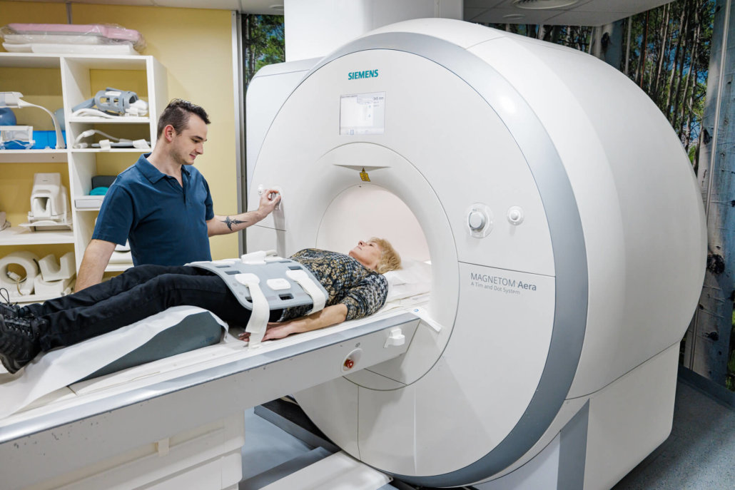
Angiography – MRI-supported
MR angiography can better diagnose certain vascular diseases. In this context, imaging allows vascular representations as well as the reproduction of cycles of blood and fluid flow within the vessels. In the meantime, in addition to the “normal” MRI procedures, there are also contrast-free blood flow images for this purpose using time-of-flight MR angiography (TOF angiography).
approx. 15-30 min
Duration of the examination
approx. 60 min
Stay in the practice
Where can you have an MR angiography performed in Munich?
MR angiography can be used to clarify various clinical pictures accurately and in a well-founded manner. Our team at each site will provide images and series of images of the areas of the vessels and discuss them with you. Your attending physician can evaluate the image material afterwards and use it for an optimal therapy.

What exactly is angiography?
This is a radiological examination that makes vessels visible (also with the help of contrast media) by means of X-ray, MR tomography or computer tomography. The images taken are displayed in the so-called angiogram. Depending on the type of vessel, angiographies are divided into three different types:
When is MRI used for angiography?
MR angiography is a gentle examination for imaging arterial and venous vessels without the use of X-rays. The previously necessary insertion of catheters into the veins and arteries is no longer necessary. This makes vascular injury, bleeding and embolisms a thing of the past!
The assessment of the condition of the vessels in the recorded body region is carried out together with one of our specialists after the examination. Vasculitis, aneurysms, smoker’s legs or renal artery stenosis can be diagnosed very well. Digital, saved versions are available for subsequent treatments.
Fields of application of MR angiographies
Coronary angiography – images of the heart
Image-guided diagnosis of heart disease takes a high priority in the examinations. First, the coronary arteries are examined. As a result of heart disease or infarction, changes or occlusions may occur. These are potentially life-threatening and must therefore be clarified safely.
The same applies to the heart chambers and their interiors. By showing their size and functional processes, any problems can be identified at an early stage. Subsequent therapy can thus be carried out in a targeted manner and under the least possible stress.
The cerebral angiography
The brain has many vessels and is highly dependent on blood supply. Malfunctions here can have life-threatening consequences. There are many causes for these disorders – from brain tumors to brain hemorrhages to vasoconstrictions. An interruption or undersupply due to blockages in the supplying vessels, for example in the neck region, can be made visible with a cerebral angiography.
Fluorescence angiography – finding retinal disorders
The eye has countless small, fine blood vessels that are essential for its function. Especially the retina (macula) can cause problems. In a so-called macular degeneration, the function of the retina is strongly affected by narrowed or clogged vessels.
The retinal disorder is diagnosed using a green dye. This fluorescein glows within the vessels under the MRI to visualize the problem areas. This eliminates the need for an additional contrast agent.
Arteriography of the legs
Angiography of the leg and pelvic arteries focuses on narrowings that can occur due to diabetes or other diseases. If, on the other hand, the veins are examined, the aim is to clarify suspected cases of thrombosis or to provide the basis for therapy of varicose veins.
Special feature: In case of intolerance of the contrast medium, carbon dioxide can also be used as a “substitute” for angiographies of the legs.
What do you need to know before the treatment?
In order to avoid a possible reaction to the contrast medium used, we recommend not eating for about four hours before the examination. On the other hand, you should drink enough water. This also reduces any possible discomfort.
Immediately before the examination, you must put down all objects that contain metal. Dentures, jewelry, hearing aids and glasses should be left in the locker room, as should watches and purses. Chip and check cards must also not be allowed near the MRI machine, otherwise the magnetic strip will be affected and the card will become unusable.
If you have metal shards in your body, be sure to say so before the exam. Especially splinters near the eye, lungs or nerve cells can move away from their previous position due to the magnetic field and thus pose a risk to your health. Our specialists will be happy to advise you in advance about the feasibility of an MRI for metal fragments in the body.
What is the procedure for angiography?
Prior to MR angiography, one of our physicians will have a consultation with you. We will inform you about the risks, the exact procedure of the examination and the benefits. Normally, we measure the blood values and clarify possible allergies to the contrast media used.
Subsequently, the contrast medium is injected into the arm arteries or the arm vein. After that, the agent must be distributed in the vascular system. Once this is done, MRI scans are performed. Due to the noise exposure, our patients are usually given hearing protection with music – this lowers the stress level during admission on the one hand and reduces any anxiety that may arise on the other.
Imaging using the time-of-flight MR angiography (TOF angiography) technique does not use a contrast agent. In TOF-MR, the freshly inflowing blood is magnetized. The resulting flow images are very accurate.
What are the risks of MR angiography?
MR angiography is considered to be relatively free of complications. The administration of the contrast medium may cause a feeling of warmth or an unpleasant taste in the oral cavity. Usually, these perceptions disappear immediately after the injection.
Rarely, patients develop an allergic reaction or intolerance to the contrast agents. These problem cases can almost always be eliminated in advance. Reasons for intolerance are often renal insufficiency and hypothyroidism. As soon as there is a potential intolerance, the contrast medium is not used.
What do you need to keep in mind after MR angiography?
After MR angiography, rest should be the first priority. Drinking plenty of fluids to flush out the contrast agent as quickly as possible is very important. In addition, the body needs a little rest. Therefore, you should refrain from hard physical work for a few days.
If inflammation or swelling occurs at the injection site, please see a doctor as soon as possible. The same applies to the occurrence of nausea, headache, palpitations or dizziness!
MRI
More about magnetic resonance imaging as an examination
CT angiography
Interesting facts about CT-guided vascular diagnostics
Angiography
Back to the overview of diagnostics by angiography


