Heart problems made visible with MRI
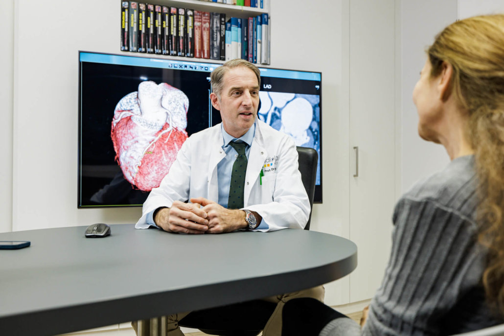
Heart problems can have very far-reaching consequences. A cardio MRI can produce very good moving images and 3D representations of the heart vessels. The method has a low impact, both in terms of examination time and radiation exposure. According to this, conclusions about certain clinical pictures can be drawn with relative certainty.
approx. 30-60 min
Duration of the examination
12 h
no coffee before the examination
4 h
Be sober before MRI
Where can you have a cardio MRI performed in Munich?
Our team of specialists performs several hundred MRIs of the heart per year. After an introductory discussion about the procedure, the risks and the necessary guidelines for preparation, the examination itself is very quick and uncomplicated. Just ask for an appointment at a location near you!

When is a cardiac MRI necessary?
Thanks to state-of-the-art equipment and experienced diagnosticians, the team at Radiologie München can also examine complex anatomical structures on the heart according to their function. With the images generated, subsequent therapeutic approaches are much more selective, less stressful and more successful. The most common areas of investigation for cardio MRI include:
MRI diagnosis of coronary stenosis
Narrowing of the coronary arteries often causes blood flow problems to the heart muscle. When the heart is placed under stress, these problems can be visualized very well. The defective blood flow (also called myocardial ischaemia) can be precisely depicted using cardio MRI.
Compared to nuclear medicine and cardiac catheterization, MRI has great advantages. It is more accurate than nuclear imaging using scintigraphy, and it is significantly less stressful than diagnosis via cardiac catheterisation. In most cases, the results of cardio MRI form a very good basis for further therapeutic approaches or necessary surgical interventions.
Inflammation & heart defects – fast help with high-resolution images
The diagnosis and treatment of myocarditis or pericarditis is nowadays hardly conceivable without imaging via MRI. The cardio MRI can determine the inflammation foci in the heart muscle or pericardium very precisely and make their distribution visible. After a few months, the progress of the therapy can then be compared via imaging. This means that therapeutic measures can be adapted exactly to the needs of the patients.
The same applies to not yet clarified functional disorders of the heart or its change in mass or size. The so-called cardiomyopathies are easily diagnosed with cardio MRI. The same applies to congenital heart defects.
Infarct treatment – focused thanks to cardio MRI
Patients who have had a heart attack are often faced with the question of which parts of the vessels can be saved (with a catheter, stent or bypass) and which are lost. Occlusion of a coronary artery almost always involves loss of myocardial tissue.
By examining the blood flow using cardio MRI, we can quickly identify what can still be saved. Even infarcts without prior diagnosis, the so-called “silent” infarcts, can be precisely clarified in terms of their effect on the arteries.
What do you need to know before the treatment?
There is little to consider when preparing for a cardio MRI. Fasting is not mandatory, although you should avoid eating or drinking for the four hours before the examination.
But what to pay very close attention to: 12 hours before the MRI NO coffee, tea or chocolate! The substances contained (so-called xanthine derivatives such as caffeine or teein) block the effect of the drug adenosine. Even in “light” coffee or the decaffeinated variety, sufficient quantities are present to affect the result. The stimulating effect that normally occurs is absent and existing damage remains invisible in the images.
It is also very valuable to have any previous findings from cardiac catheterization, echocardiography, or scintigraphy with you for the exam. If you are taking theophylline and nitro preparations, these should be suspended for 24 h in consultation with your treating physician.
If you suffer from claustrophobia or other anxiety, it is appropriate if you do not come to the MRI alone. Due to the possibly necessary administration of a sedative, you are not allowed to drive a motor vehicle, and normally you are not even allowed to actively move in road traffic (no cycling or walking longer distances) for insurance reasons.
If necessary, shaving of the corresponding attachment points for the ECG electrodes may be required if there is a lot of body hair on the chest.
What is the procedure for a cardio MRI?
At the beginning of each examination, our specialists conduct a personal informative talk with the patient. This includes the exact question, possible risks during the examination and also the exact procedure of the cardio MRI.
Following this, the drug adenosine is injected during a stress MRI. Its effect and composition is also part of the preliminary conversation. The additional contrast medium does not contain iodine, unlike the usual CT media, and is therefore safe for patients with thyroid disorders. It may be necessary to give an additional sedative to alleviate anxiety before the examination. Of course, this is done under permanent medical supervision.
The examination itself usually takes between half an hour and an hour. There will be loud knocking in the MRI. This is due to the solenoids on the unit switching on and off quickly and is a normal thing. For a more relaxed examination, headphones are used to significantly mitigate this influence. Through them you will also receive instructions again and again, mainly for holding your breath for a short time. This is necessary to maximize the sharpness of the images. However, this will not be necessary for longer than 15 seconds per image creation.
Directly afterwards, one of our experts will talk to you about the results and you will receive your own report CD with the images created.
Cardiac CT
Spatial & temporal imaging using CT
Coronary calcium measurement
Detection of calcifications of the coronary arteries
Cardiac diagnostics
Back to the overview of cardiac diagnostics

