Breast ultrasound
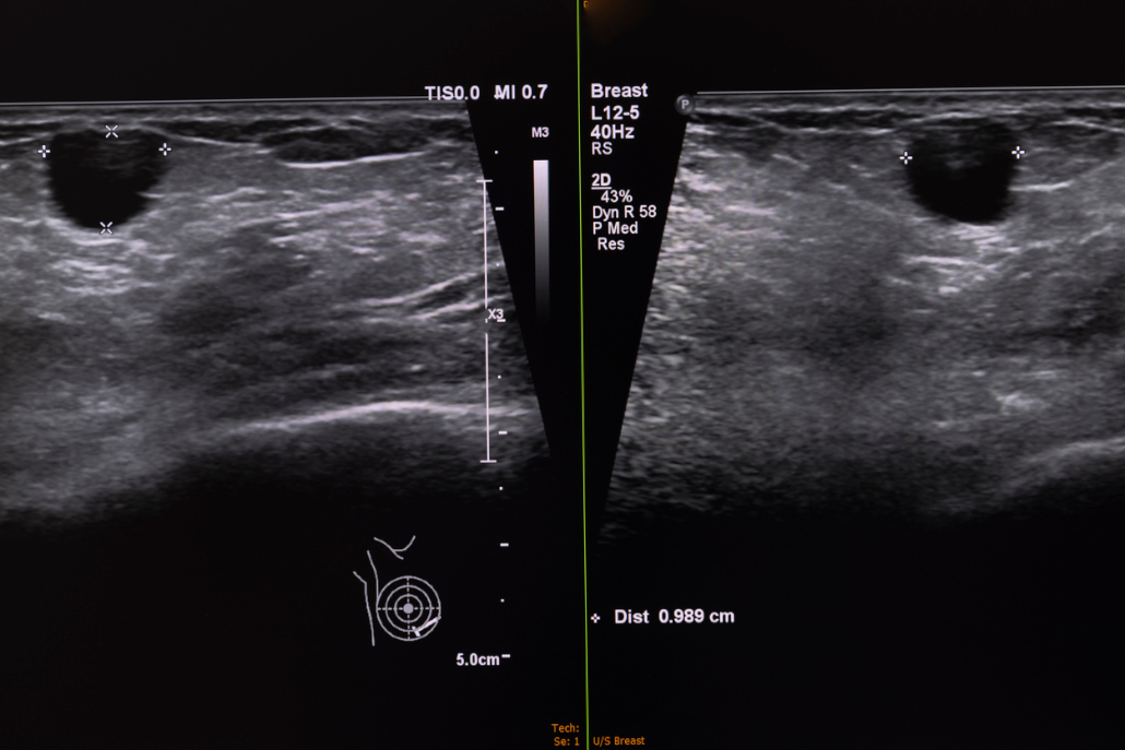
For the examination of the female breast, ultrasound with high frequencies up to 18 MHz is used in mammasonography. Ultrasound is an excellent method to increase the validity of mammography. Especially in dense tissue, this improves the detection of breast cancer.
approx. 15 min
Duration of examination
none
Known side effects
Where can you have a breast ultrasound examination in Munich?
Especially in the case of dense breast tissue, this supplements the informative value of a mammogram. Simply ask for an appointment with one of our specialists at Radiologie München.

How does a mammary sonography work?
In mammary sonography, the breast is examined without radiation. The ultrasound probe is used to “scan” the breast and armpit. With the help of ultrasound and corresponding physical laws, a film is created on the monitor, which is composed of continuous images. These images are ultimately sectional images in different planes.
- exact localization of the tumor
- Differentiation of fluids (cysts) and solid (solid) nodules
- a reduction in false-positive findings and false-negative findings in addition to mammography
When is an ultrasound examination needed?
Women up to the age of 30, pregnant and breastfeeding women can feel a lump in the breast themselves. Sometimes the gynecologist also finds an abnormal palpation. If palpation findings are conspicuous, an ultrasound examination can first be performed in these women.
Mammary sonography leads to quickly available results and can be discussed with the patient directly after the examination.
Is there anything to consider before mammasonography?
Special preparation by the patient is not necessary. You should remove neck jewelry during the examination. Breast piercings or body jewelry in the breast area must be removed beforehand.
Are there any side effects?
During mammasonography, you may experience slight pressure sensations from the transducer on the breast being examined. There are no subsequent side effects.


