Link to: Angst im MRT
MRI without claustrophobia
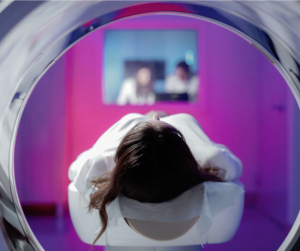
Claustrophobia in a narrow tube is not an uncommon problem, even among our patients. That’s why at many locations we only use equipment that has extra-large diameters and narrow equipment depths. In addition, our staff is trained in dealing with patients with claustrophobia.
Technical equipment
At our various locations, we work exclusively with MRI equipment from Siemens-Healthineers, formerly Healthcare. With this optimal equipment, we can use the most effective imaging for accurate diagnoses with the least possible stress for our patients.
Where can you have an MRI examination in Munich?
You can have various MRI examinations performed in our group of practices. In addition to joint MRI and vascular MRI, we also offer organ MRI for the heart, kidney or gastrointestinal tract. Special diagnostics for cancers such as prostate or breast cancer are an integral part of our daily radiological routine.

1
2
3
4
5
6
7
8
9
1
2
3
4
5
6
7
8
9
How does an MRI scan work?
Magnetic resonance imaging (MRI, magnetic resonance imaging) works by using magnetic fields and radio waves. No X-rays are used. The patients are therefore not exposed to radiation during the MRI examination.
Radiation free examination
X-ray examinations use radiation to shine through body tissue and make certain parts visible. With an MRI, on the other hand, the device uses the physical properties of the body’s cell nuclei. Like all atomic nuclei, those in the body rotate on their own axis. This “nuclear spin” leads – similar to a bicycle dynamo – to the formation of a minimal magnetic field. The alignment of this field is usually purely random. If a strong magnetic field is now generated around the body, the “mini magnetic fields” of the individual atoms align themselves – in the longitudinal direction of the human body.
So much for the physical basis – the MRI generates a strong magnetic field. In this field, which is completely harmless to humans, the MRI machine now emits radio waves that penetrate the human body and thus change the alignment of the atomic nuclei, i.e. deliberately “mess them up”. Since atomic nuclei quickly return to longitudinal alignment after the radio wave pulse, these motions can be measured.
Differentiation between sick and healthy thanks to magnetic field evaluation
The water atomic nuclei that are most abundant in the body are particularly important. Their frequency varies depending on the nature of the tissue. And so measurably different values result, which the system then assembles into evaluable images. The images then not only show the different areas of the body region examined, but also allow a good delineation between healthy and diseased tissue.
Who is NOT allowed to have an MRI?
Due to the magnetic fields, patients with pacemakers and other stimulators are not allowed to undergo an MRI, or only under very special conditions. Prior consultation with our medical specialists is absolutely necessary!
In principle, the exclusion applies to carriers of:
- Pacemakers,
- Cochlear implants,
- Neurostimulators and
- Insulin pumps.
Shrapnel or other metal fragments in the body are also critical. Please be sure to tell our medical team if you are carrying such foreign bodies. Endoprostheses for the hip and knee, stents, dental fillings and bridges etc. are not problematic. Ask in the preliminary consultation and our specialists will clarify the corresponding problems.

What should be considered before the MRI examination?
You should arrive at the registration desk approximately 20 minutes prior to the start of the examination. No special preparation is necessary for most examinations. In particular, you do not have to be sober. Exception: For vascular and organ MRI, such as the upper abdominal organs, liver, gall bladder, or pancreas, you should be sober! In this case, you are not allowed to eat or drink anything after 10 p.m. on the previous day. For all other MRI examinations, you can eat normally before and after the examination.
Can I take my medication before an MR scan?
Medications can basically continue to be taken, according to your normal medication regimen. Relevant interactions with the contrast agents we use are not known to date. Vor der MRT sollten Sie Ihre dauerhaft genommenen Medikamente mit unseren Fachärzt:innen absprechen.
What should you wear for an MRI scan?
In general, we recommend wearing comfortable clothes that you feel good in. At the same time, try to avoid any kind of metal in the clothes. For chest MRI, shoulder MRI, and pelvic area MRI, as well as for spine exams, you should not wear clothing or underwear with metal hooks or metal fasteners, or remove them in the locker room before the exam. Trousers with metal buttons, zips, press studs or belts can also have a negative effect on image quality and should be removed before the examination.

Problem body jewelry and makeup!
All metal-containing items such as hair clips, earrings, jewelry, and piercings must be removed prior to the examination. If piercings are not removable, they must be shown in detail to the examining personnel to enable a clear classification of this source of error. Furthermore, examinations of the head and neck area may be affected by makeup, as some products contain minimal amounts of iron. Please inform our staff already before the examination if you wear “permanent make-up”.
What should you bring to the examination?
If you are to have an examination with contrast injection, we need the so-called creatinine value. This is a laboratory value that can be determined for you in your GP or specialist practice. Ideally, the value should not be older than 4 weeks. However, if you have no pre-existing kidney disease, older values may be sufficient. If in doubt, you should ask at the appointment desk. If available, you should bring documents on allergies (allergy passport) and implants.
MRI and pregnancy – does it fit?
 Can I have an MRI during pregnancy?
In principle, there are no known harmful effects for the unborn child. Nevertheless, MRI examinations are excluded in the first third of pregnancy (1st trimester). Exceptionally, however, this would be feasible in the case of a health risk to the mother (a so-called vital indication). In the later stages of pregnancy, MRIs can be scheduled and performed without problems.
Can I breastfeed after an MRI?
After birth, the question of breast milk impairment often arises. To reassure mothers, we recommend that breast milk be pumped and discarded during the first 24 hours after MRI.
In general, minimal amounts of contrast medium may pass into breast milk when contrast medium is administered. However, the infant cannot absorb this through the intestine. So there is no danger.
Is an MRI dangerous to health?
In the course of the examination with MRI, the issue is often whether health problems occur after an MRI. Clear answer: No! So far, no harmful effects are known. The tissue heating caused by the magnetic field is minimal and below legal limits. These limits are so low that even if they were reached, no permanent damage would occur.
The loud noises in the MRI can be stressful – but you will receive hearing protection against them in any case, either via headphones or earplugs.
 Is there any radiation?
Magnetic resonance imaging does not use ionizing radiation, unlike conventional X-ray examinations or computed tomography. Therefore, they are not exposed to radiation or radioactivity.
Can children be examined in MRI?
Yes. In many cases, magnetic resonance imaging is the method of first choice in the diagnosis of pediatric diseases. Since the child’s body is particularly sensitive to radiation, exposure to X-rays and CT should be avoided as far as possible.
Minor patients must be accompanied by a parent or guardian for MRI. During the preceding clarification, the consent with signature is also necessary – this can mostly not be done by the minor.
Adolescents can bring a written declaration of consent from a parent or guardian as a substitute, but this must be agreed in advance by telephone and, if necessary, clarification must be provided. Please contact us if you have any questions about this.
How does an MRI work?
Magnetic resonance imaging can take between 10 and 30 minutes. Normally, you lie on your back and the body region to be examined is moved into the large, ring-shaped opening of the device.
What to consider during the examination:
- Lie as still as possible
- Even breathing
- Brief breath holding may be necessary on instruction
During the examination, our medical staff will monitor you via a screen or directly from the machine.
When an MRI scan is running, loud knocking noises occur. These are part of normal operation and are caused by the generation of the rapidly changing magnetic fields. You will be provided with hearing protection for a comfortable examination.
Use of an intravenous contrast agent
When examining very similar tissues or tissue changes, the use of an intravenous contrast agent is necessary. Otherwise, it would not be possible to differentiate between diseased and healthy tissue areas, or only with difficulty. In addition, an intravenous contrast agent helps in the (early) detection of tumors or inflammatory foci.
The contrast agent is applied via the arm vein during MRI. The contrast media used are generally well tolerated and non-radioactive. Within a few hours it is completely flushed out of the body with the urine. If you suffer from certain problems, such as kidney disease, which affect tolerance, clarify this together with our team during the preliminary discussion.
Link to: Aktuelles