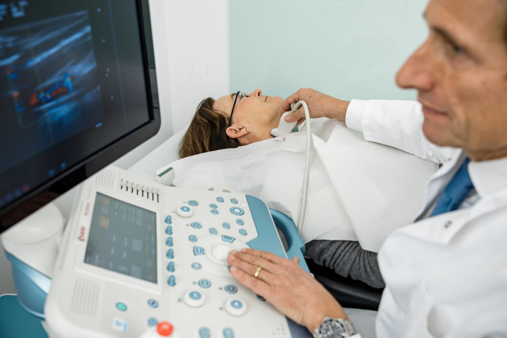
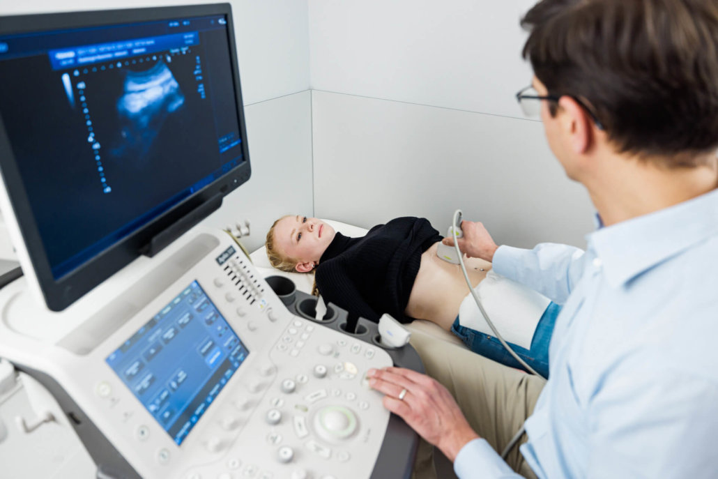
View with ultrasound
Sonography, i.e. imaging by ultrasound, is used in many ways at Radiologie München. In addition to abdominal and soft tissue examinations, ultrasound images are particularly used in breast diagnostics and thyroid cancer detection. Particularly in the latter, sonography also allows for the visible puncture of any tissue areas.
approx. 10-20 min
Duration of the examination
approx. 45 min
Stay in the practice
Your sonography at Radiologie München
We offer ultrasound examinations at all of our radiology locations. You can rely on our competent sonography experts, who will explain the results to you after the examination and make the findings available digitally on the patient platform.

How does a sonography work?
The method originally developed from sonar technology for locating ships and is based on the application of high-frequency, mechanical vibrations. The radiation-free, completely harmless and painless ultrasound waves are reflected by different tissues to varying degrees – thus creating an echo that is subsequently recorded on the transducer.
This echo is converted into an image called a sonogram. In this way, we can see many regions of the body. Thus, the procedure is suitable for the initial clarification of complaints or for the first exclusion of pathological organ changes.
Where does sonography offer high diagnostic potential?
Sonography is an important procedure in the differential diagnosis of an acute abdomen, gallstones, or the evaluation of vessels, particularly with regard to stenoses in the neck and extremity arteries. It is also suitable for examination:
- of the thyroid gland,
- of the heart,
- of the kidneys,
- of the urinary tract,
- the urinary bladder and
- during breast diagnostics
However, it is not uncommon for sonographic findings to require further clarification by follow-up examinations such as MRI, CT, or PET/CT.
What should you consider before the examination?
In ultrasound examinations of the abdomen, the image quality is highly dependent on the contents of the bowel. We therefore recommend avoiding flatulent foods the day before sonography. On the day of the examination itself, please appear as fast as possible. You should not consume food or fluids until sonography.
For examinations of the neck, breast or connective tissue, no restrictions on eating or drinking are necessary beforehand.
Sonography usually takes between 10 and 20 minutes. After a short discussion in our practice, the examinations are then performed lying down, in some cases also sitting up. To guarantee perfect imaging, a neutral gel is applied to the transducer of the examination device. This prevents the contact distance between the transducer and the skin from breaking. The gel is usually highly compatible and can be removed without residue after sonography.
During sonography, one of our specialists moves the transducer over the corresponding body region and examines this area from different echo positions. The displayed images are digitally archived and linked to your patient file. After the examination, our doctors will not only show you the sonograms, but will also explain the findings to you. Based on this, they will also recommend possible further forms of diagnosis in case the result is not clear.


