What is brachytherapy?
What is
brachytherapy?
Brachytherapy stands for radiation therapy in which an enclosed, radioactive source is introduced into the body. In doing so, the radiation therapists store this source either directly in the target area of irradiation or in its immediate vicinity. Other names for brachytherapy are: “internal radiotherapy” or “irradiation from the inside”.
Use for which tumors?
The types of tumors treated with brachytherapy are diverse. Mostly the following cancers are treated:
Where is brachytherapy available in Munich?
The treatment of patients by means of brachytherapy can be carried out by our team in Munich-Schwabing. We support colleagues from other specialties in the development of an individual treatment strategy with the aim of achieving the lowest possible burden while at the same time providing optimal therapy. We offer ONLY intracavitary brachytherapy (e.g., for uterine cancer) in our practice.

What are the types and differences?
Brachytherapy is divided into two forms:
- Intracavitary brachytherapy and
- Interstitial brachytherapy
Intracavitary brachytherapy
Intracavitary brachytherapy stands for the use of body orifices and body cavities for positioning the radiation source. The radiotherapist inserts it in such a way that it is placed directly at the target region with the tumor to be treated. For tumors close to the surface, the radiation source can also be applied directly to the skin (contact brachytherapy).
Interstitial brachytherapy
In interstitial brachytherapy, small radiation sources (seeds) are introduced directly into the tumor tissue by means of cannulas and/or catheters. The seeds deliver their radiation dose there over a longer period of time. The most common application of this procedure is the treatment of prostate cancer.
Advantages and risks of brachytherapy
The advantages of brachytherapy lie in the special mode of action of this form of therapy. Due to the narrowly limited area of irradiation to the source, the surrounding organs and body area are spared. This reduces the risk of side effects and simplifies the management of side effects.
In addition, only a few treatments are necessary due to the comparatively high radiation doses. These sessions can mostly be done on an outpatient basis – a considerable relief for our patients.
Procedure of brachytherapy
Brachytherapy starts with a thorough, medical examination. All diagnostic results (MRI, CT, etc.) are used to precisely determine the location of the tumor area to be irradiated. The risk of damage to neighboring structures is also assessed in this way. The optimal position of the applicator or the irradiation sources can be planned.
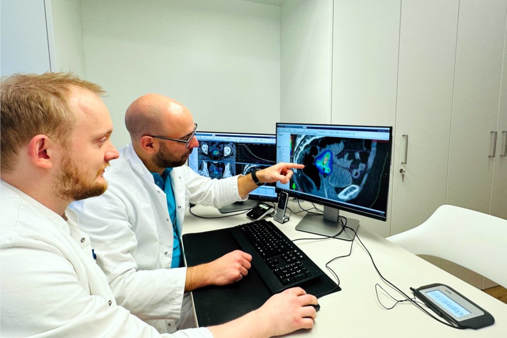
After placement of the applicator, our radiation therapist(s) recheck the correct position with CT/X-ray, MRI and/or sonography. Medical physicists and radiotherapeutic specialists plan the exact course of the radiation treatment together with the help of therapy planning software. The individual radiation plan is optimally tailored to the patient’s individual anatomy.
During the actual treatment, the radioactive radiation sources are introduced into the applicator under computer control. According to the treatment strategy, the dwell time of the radiation sources was calculated in advance and now implemented accordingly. After administering the prescribed dose, the radioactive source retracts. The applicator is removed.
What side effects may occur?
Depending on the risk and degree of side effects, a distinction is made between possible acute and delayed effects:
In the case of acute side effects, short-term irritation of the skin or mucous membranes may occur in the area directly adjacent to the radiation source. This may also manifest as more frequent bowel movements or diarrhea. After the end of treatment, these irritations usually subside completely.
So-called. Late side effects occur with permanent damage to adjacent organs and tissue parts. These are very rare and mostly mild. Mainly, scar tissue formation may occur in the treatment area. Vaginal therapy for uterine carcinoma, for example, may result in vaginal stenosis.


