Main advantage of tomosynthesis – the earlier breast cancer is detected, the better the treatment options. Imaging mammography has previously relied on 2D imaging. Tomosynthesis is used to produce three-dimensional images of the breast. This significantly increases the quality of diagnostic imaging, as overlapping tissue areas are more accurately visualized.
approx. 30 %
higher tumor detection
approx. 30 %
Decrease in false positive findings
Where can you have tomosynthesis performed in Munich?
The innovative three-dimensional imaging (3D) of the breast using tomosynthesis is one of the most modern methods of early breast cancer detection in Germany. Particularly in the case of dense breast tissue, this enables clear diagnoses to be made. Simply ask for an appointment with one of our specialists at Radiologie München.

How does a tomosynthesis work?
As with a digital mammogram, tomosynthesis uses X-rays to view the breast. In the process, the X-ray tube swivels up to 50 degrees around the chest. The slice images taken in this process are converted into a 3D model of the examined breast by means of the software technology used. The advantages are especially in dense breast tissue with
- a millimeter-precise localization of the tumor,
- a significantly more sensitive presentation (20-30% increase in tumour detection),
- a reduction in false-positive findings, and
- a blanking of the overlying glandular tissue
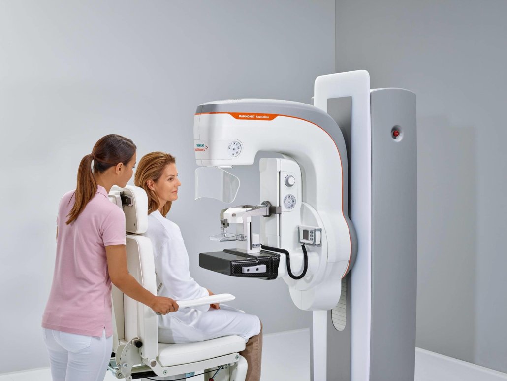
Siemens Healthineers Mammomat Revelation
Technology made in Germany for optimal examination results: the Mammomat Revelation tomosynthesis device from Siemens used by our specialists is a system that automatically determines breast density. Thus, in addition to the potential tumors to be examined, their margins are also optimally delineated. The accuracy of the localization is about one millimeter. Precise localization is important for the subsequent treatment of the tumor.
When is tomosynthesis needed?
Palpation examination
It is recommended that women over the age of 30 have a check-up for breast cancer once a year at a gynaecological practice. For this, the lymph nodes in the area of the armpits, the breast and collarbone are palpated. The size and shape of the nipples are also checked, looking for any changes or deformities.
In addition, check for fluid leakage when the nipple is gently pressed. If there are palpable findings or if the situation is unclear due to dense breast tissue, a precise diagnosis can be made by means of tomosynthesis.
3D mammography
The layer-by-layer X-ray examination takes only a few seconds. For the duration of the images, the breast is compressed using a Plexiglas compression clamp. Many women find this unpleasant, in very rare cases even painful, but this method provides a special clarity to the images taken. Assembling the images to a 3D model is done directly after the acquisition. Our specialists then discuss the diagnosis based on this with the respective patient.
What should be considered before a 3D mammography?
On the day of the examination, we advise avoiding body lotion or deodorant. The particles contained therein can be displayed with the acquired images and thus falsify the diagnostics. You should remove ear and neck jewelry during the examination. Breast piercings or body jewelry in the breast area must be removed beforehand.
Are there any side effects?
During the examination, there may be a sensation of pressure on the breast being examined. The plexiglass discs used are adapted to the ergonomics of the female body, but the fixation of the breast can be perceived as uncomfortable. Follow-up side effects are absolutely rare and correspond to those of normal X-rays. Radiation exposure is about the same as with 2D digital mammography.


