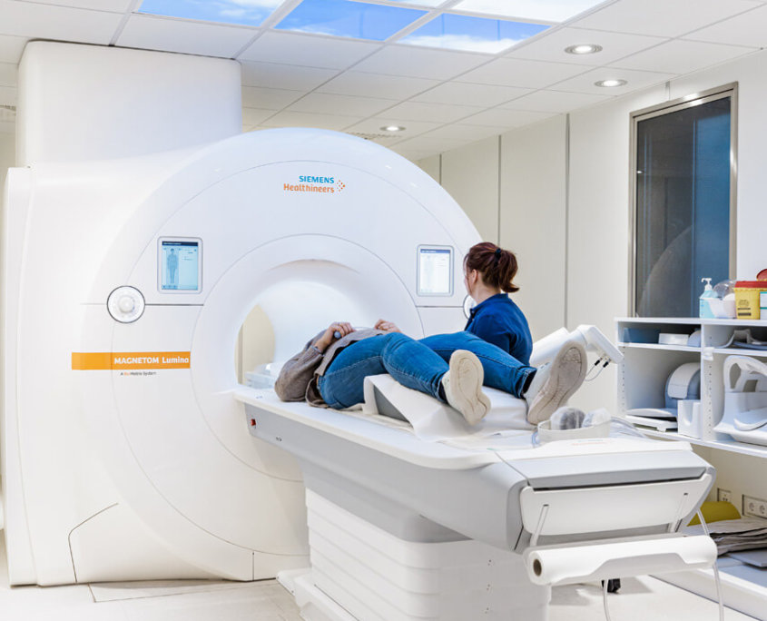
Breast cancer diagnosis covered in the best possible way
Breast cancer is the most common cancer diagnosis in women – MR mammography helps with early detection. Digital mammography – including 3D tomosynthesis – has already reduced the number of severe courses of breast cancer in recent years. MRI or magnetic resonance imaging of the breast (breast MRI) can offer critical advantages in early detection when mammographic and sonographic findings are unclear and in follow-up to evaluate for recurrence.
approx. 15 min
Duration of examination
> 90%
Accuracy in breast cancer tumor detection
Where can you have an MR mammography in Munich?
Our specialists in breast diagnostics offer breast MRI at several locations in Munich. Choose the location that suits you and ask for an appointment.

Statements after MR mammography
Breast MRI has proven to be one of the imaging modalities for the diagnosis of breast cancer. The examination is performed in the magnetic field using a special mammary coil in the prone position. Radiation exposure (X-rays) does not occur. After administration of a contrast agent (rare earth, gadolinium), tissue with enhanced contrast uptake (tumor tissue) can be delineated. In this way, it is possible to assess, particularly in dense glandular tissue, whether a suspected focal finding exhibits criteria for malignancy. It is also important to know whether the focal finding is the only finding in the breast or whether, for example, there are other foci in the surrounding area or even in the other breast that cannot be seen with mammography and ultrasound.
The difference in accuracy between mammography and MRI results from the fact that MRI is not limited by the density of breast tissue, as the breast is imaged slice by slice using a cross-sectional imaging technique.
The timing of a woman’s menstrual cycle (menstruation) is important for the timing of breast MRI, as there are hormonal changes depending on the cycle. At the time of examination, it should be as “quiet” as possible in the breast, which is the case from the 9th to the 16th day of the cycle.
When is MR mammography performed?
A breast MRI can generally be used for the early detection of breast cancer. For women who have implants in their breasts, breast MRI is usually the alternative to mammography for meaningful screening.
In addition, breast MRI is often used in the following cases:
Unfortunately, there is a limitation that MRI sometimes cannot be used in patients with pacemakers or other important medical implants, or cannot be used without prior consultation.
Is there anything to consider before MR mammography?
The mammary gland tissue is subject to changing hormonal influences. Depending on the cycle-dependent hormone level, the absorption capacity of the harmless, non-iodine-containing contrast agent is altered. Since this can reduce the validity of the examination, we ideally perform the MRI from the 9th to the 16th day of the cycle. Women who are in menopause/climacteric are not affected by this time restriction.
Please bring your findings from previous examinations with you to the examination appointment. This facilitates and improves our diagnostics. Thus, previous MRI examinations, mammograms, ultrasound scans and, if applicable, reports of findings after biopsy and therapy are important to us.
Unless you have known intolerances to contrast media or a pacemaker, there are no necessary preparations. If they suffer from kidney function limitations, we need the current creatine value. Please bring this with you to the examination appointment.
You do not need to be sober for the examination. You can also take your medications as usual. If you do not need a sedative during your breast MRI, you will be ready and able to drive immediately afterwards.
If you require the administration of a sedative due to claustrophobia, please bring an accompanying person! If you have any questions, just contact our staff.
What is the procedure for an MR mammogram?
At the beginning, an in-depth discussion takes place with our specialists about the procedure, the informative value of the images produced, the effect of the contrast medium used, as well as possible fears and questions of the patient. An access is then established via the arm vein, through which the contrast medium is administered during the examination. This contrast medium does not contain iodine and is therefore uncomplicated for patients with thyroid problems.
MR mammography, unlike other MRI exams, is performed in the prone position and takes approximately 12 to 15 minutes. The breast is supported in the MRI coil for this purpose, without any pressure pain for the patient.
To achieve the best possible images, it is enormously important that you do not move during the entire examination. If you find this difficult, for example due to claustrophobia or nervousness, you can also be given a sedative. We recommend that you always have an accompanying person with you. Should we give sedatives, they will not be allowed to drive. Otherwise, in the event of an accident, your insurance coverage would be gone!
Immediately after the examination, the findings are discussed. A corresponding data CD will then be given to you as the patient.

